E. Solarte1, A. Reyes1, J. A. Montoya1, J. Arroyabe2, R. Neira3
1Grupo de Optica Cuántica, Departamento de Física
Universidad del Valle, A. A. 25360, Cali, Colombia
2Centro Internacional de Agricultura Tropical, Cali
3Centro Médico Imbanaco, Cali
SUMMARY
Recent studies show an apparent effect of small doses of the irradiation with laser light 635 nm, made immediately before liposculpture interventions, which use the tumescent technique. These effects are macroscopically affirmed by a freely fat extraction and microscopically appear as the disrupture of the cellular membrane, which facilitates the exit of fat. In order to observe the effect in separated adipocytes, dense dissolutions of adipocytes, separated by centrifugalization, were prepared in tumescence solution. The dissolutions were irradiated in vitro with laser light (Laser Diode, 635 nm). Visible light transmission spectra were studied for different dissolution concentrations and different irradiation doses.
INTRODUCTION
Clinical observations [1, 2] made from the end of 1999, presumably show that the external application of laser radiation, previous to interventions of liposuction by the tumescent technique [3] presented very interesting results. Demonstrating that the fat exit was facilitated due to an interaction between the light and the cells. The microscopic observation shown membrane pores and fat drops enter the interstitial space. At the same time, the other structure like interstitial space, connective tissue and capillary cells, are conserved. With the purpose of corroborating a possible irradiation effect on the fat distribution, image studies by MRI were performed [1].
With the purpose of explaining the mentioned effect, and to study the laser radiation pagation through the tissue, we started (2000) a research work in our quantum optics laboratory at the Universidad del Valle. Transmittance studies of visible light in dissolutions of fatty cells have been made [1, 2, 4]; the propagation of the Laser Light through adipose tissue samples has been studied [5] and experiments on laser light diffraction have been made [6] using thin adipose tissue samples. In this work the results of optical transmittance measurements in dissolutions of adipocytes are reported. Transmittance spectra were recorded for different concentration and different irradiation powers and irradiation times.
MATERIALS & METHODS
Dissolutions of centrifuged adipocyte cells are used as a reliable tool and the mixture was prepared for tumescent infiltration. Defined masses of concentrated adipocytes were placed in soft polyethylene flasks that contained given volumes of the infiltration solution. For this case, portions of 1.8 g were taken and then they were dissolved in 40 milliliter of solution. The mixture was remained to rest for 20 minutes and the exudate was extracted with a syringe to the spectrometer cuvettes. The rest time determines the dissolution concentration. A sample of infiltration solution serves as reference for the transmission spectra. The transmittance spectra were taken for the sample without exposure to laser radiation and successive spectra were recorded as it was exposed to the laser radiation during fixed consecutive exposure times by a fixed laser power. The laser used was a semi-conductor clinical laser, emitting at 635 nm, with a power of 3.55 mW.
For the spectra acquisition a miniature diffraction grid, optical fiber spectrometer system with a four-way cuvette holder was used. The include detection system was a linear array of 2048 CCD elements. The spectra were accumulated in a normal PC and they were processed later using a suitable data and graphics processing software.
Two types different types of irradiations procedures were utilized, taking advantage of the four-way cuvette holder. In a first sequence of measurement, low density dissolutions were irradiated in the cuvette, outside the spectrometer cuvette holder, placing the laser beam directly on the liquids upper surface using a laser power of 3.55 mW. The transmittance spectrum was then recorded.
On a second series, an optical fiber was used to directly irradiate dense dissolutions in the cuvette, within the camera of the spectrometer, with a power of 300 microW, observing the transmittance signal simultaneously. The second type of measurements were made with the purpose of observing the behavior of high concentration and the low radiation doses that presumably are within the radiated organism.
RESULTS
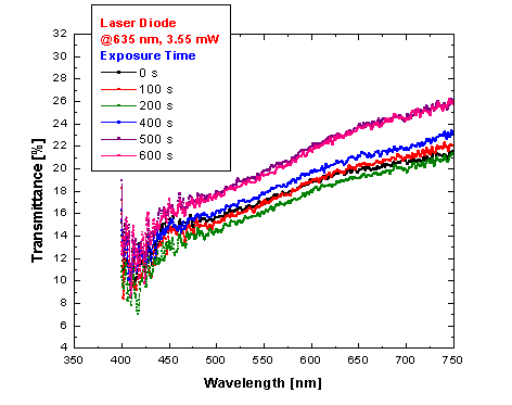
The spectra of samples directly radiated with the laser, have the form shown in Figure 1. It is observed that the transmittance increases with the irradiation time, and saturation is reached, with maximum values of about 27%. This behaviour was observed [ 1, 2, 4 ] previously in dilutions obtained from the tissue exudates, when an adipose tissue block was under hydration in the infiltration solution. All all these cases transmittance changes of about the 10% where obtained.
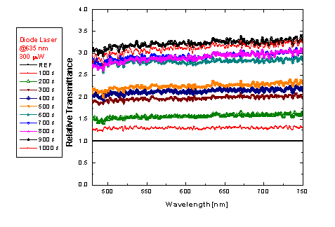
For the second type of measurements, a behavior similar to the previous one is observed, only that in this case the saturation happens in longer times. Transmittance increasing was also observed during the irradiation period. The main difference comparing with the low density result is in which great variations of the transmittance happened. To detect the changes, the ratios between the transmittance of the irradiated sample to the non-irradiated one where studied.
Figure 2 shows the relative transmittance of a sample radiated with 300 microW of laser power. The transmittance variations reach 3 times the respective value of the non-radiated samples, a change of 300% could be determined.

Figure 1. Transmittance spectra for a low dense dissolution of adipocytes, radiated directly with a laser diode at 635 nm, 3.55 mW.

Figure 2. Relative transmittance for a dense dissolution of adipocytes, laser radiated with an optical fiber, about 635 nm, 300 microW.
DISCUSSION
From the physical point of view, it is to consider that the adipocytes are great cells and of almost spherical form of 100 microns of diameter. Scattering or dispersion effects and absorption can explain the dissolution opacity or turbidity. For the used wavelengths (0.4 to 0.85 microns) the effects of Mie scattering are predominant on those of Rayleigh scatter and therefore the amount. The form and size of the centers of radiation scatterers defines the behavior of the light. If now, by some process, the form or the number of mean points of impact is altered, these alterations appreciably modify the distribution of the light and therefore the average transmittance. Transmission spectroscopy observations indicate that when irradiating the samples, the transmittance increased denoting the variation in the number or the form of the scatterers exist. These observations are consistant with the ones made by electronic microscopy that show morphological changes of the adipocytes induced by irradiation.
CONCLUSION
The results obtained by transmission spectroscopy allow observing an effect of the irradiation adipose cells, which is translated in changes of the optical transmittance of the irradiated dissolutions, measured in relation to the transmittance of the non-radiated dissolutions. Variations until of 300% in the transmittance were observed for dense dilutions and low radiation doses. This fact is an indicator of variations in the size, the form or the number of radiation scatterers.
ACKNOWLEDGEMENTS
The authors (ESR, MAR, JAM) thank to the University of the Valley for the support to make this work and to attend the CNF. All the authors thank to the CIAT and the Medical Center Imbanaco for the collaboration for this project, and COLCIENCIAS for the financial support under the 1106-05-10107 project.
REFERENCES
1. R. Neira, J. Arroyave, H. Ramírez, C. Ortiz, Solarte, F. Sequeda, M. I. Gutiérrez. Low Laser Level Assisted Lipoplasty: To new Technique., Presented at the World Congress on Liposuction Surgery, Michigan, October 2000.
2. R. Neira, C. Ortiz, J. Arroyabe, H. Ramírez, M.i. Gutiérrez, To Reyes, And Solarte. 2000. Procc. Vii National Optics Meeting. Sept. 25 -29, 2000, Armenia, Colombia. To be published.
3. J. To Klein. Tumescent Technique. Amj Cosmet Surg 4, 263, (1987).
4. R. Neira, J. Arroyabe, M. To Reyes, And Solarte. Irradiation effects on Adipose cell Dilutions.. Personal for publication to Laser Med. Surg. (2001).
5. M.s To Reyes, F. Rebolledo, J. To Montoya, C. Ortiz, And Solarte, R. Neira. Dispersión de Coherent Luz by Fatty Weave samples. Personal for presentation to XIX the CNF, Manizales, September 2001.
6. To F. Rebolledo, M. To Reyes, J. To Montoya, And Solarte. Difracción and Dispersion of Coherent Light by random difractores and their application to the dispersion by Fatty Weave. Personal for presentation to XIX the CNF, Manizales, September 2001.
